Surgery Day
What You Should Know About Mohs Surgery
Mohs micrographic surgery is a specialized technique for the total removal of skin cancers. The procedure is done in several steps:
I. A local anesthetic (usually Lidocaine) is injected around the skin cancer.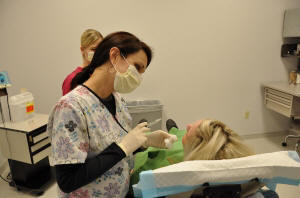
2. Surgical removal of the cancer is done in a specific fashion so that the entire undersurface and skin edges can be examined microscopically.
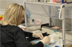
3. The tissue is dyed and a corresponding map is drawn so that any tumor found microscopically can be located exactly on the patient.
4. The surgeon then examines the slides under the microscope to look for cancer cells,
5. Subsequent tissue is removed in a similar manner until no more cancer cells are found. In most cases, the surgery is done on an outpatient basis; however, in some cases, the doctor may recommend hospital admission. This is especially true for patients with large tumors, but admission is the exception rather than the rule.
How to Prepare for Surgery
 No real preparation, other than a good night’s sleep, is required. Eat a light breakfast on the day of surgery. If you are currently taking medication, continue as usual unless directed otherwise by your physician. Eliminate any non-physician prescribed aspirin for at least two weeks before surgery. This is because aspirin tends to prolong bleeding during the operation. Also, eliminate medications containing ibuprofen, commonly found in Advil, Aleve and Motrin, for at least three days before surgery. If you need a pain reliever, you may take acetaminophen, which is found in Tylenol.
It is best to wear a shirt that buttons down the front. No makeup or jewelry should be worn if surgery is to be performed on your face. Bring a good book or magazine with you, as you will spend a significant amount of time waiting while the microscopic slides are prepared and interpreted.
What to Expect on the Day of the Surgery
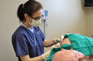
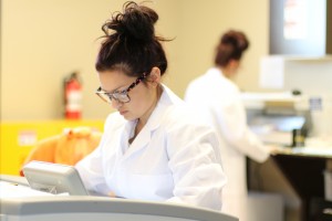 Shortly after your arrival, you will be taken to one of the treatment rooms, where the tumor area will be numbed with a needle. The doctor will remove a thin layer of skin surrounding the cancer. After this has been done, any bleeding will be stopped with an electric cautery machine. You will then be bandaged and be able to return to the waiting room. By this time, the removed tissue will be in our laboratory. There, it is cut, dyed and made into microscopic slides. It usually takes 20-30 minutes for the layer of tissue to be removed and the bleeding to stop. However, it takes about I hour for the tissue to be prepared into microscopic slides for examination. During this time you may chat with the person accompanying you, read a book or step out for fresh air.
 If examination of the microscopic slides reveals that your tissue still contains tumor cells, the procedure will be repeated. Further tissue is removed only from the areas where tumor cells were found. The goal is to remove all of the skin cancer while preserving the greatest amount of healthy tissue. However, skin cancers can grow deeply and develop roots extending beyond the area that you actually see. As a result, the final size of the surgical incision will be determined by the extent of the tumor.
The average number of surgical sessions required is one to three. However, you may require more before your skin cancer is completely removed. Fortunately, this can usually be done in the course of a single day. When surgery is completed, a decision will be made as to the way to manage your wound.